Taylor West – “Digital Redisplay at the Oxford University Museum of Natural History”
Digitization – the act of creating digital, virtual models of existing museum objects and displays – within the museum space can sometimes be a contentious topic. Curators and museum professionals champion the immersive experience that accompanies a traditional in-person visit, as many exhibits rely on more senses than just sight to nurture their narratives. Digitization can seem threatening to this experience, as some may assume that it promotes virtual exhibition at the cost of in-person visits.
It is important, however, to consider the broad array of positive outcomes that digitization of museum objects and exhibits can provide. Digital surrogates improve accessibility for those who may not have the means or abilities to visit a museum in-person. Digitization may enhance a visitor’s experience through accompanying interactive displays, as one can closely inspect an object otherwise locked behind a glass case. Digital displays can also offer more information than what may be presented on the accompanying text description for an object, allowing for improved learning.
Earlier this year, I traveled to the Oxford University Museum of Natural History (OUMNH) to complete my Museum Studies internship. My goal was to work with museum staff to create and implement a “digital redisplay” in parallel with their ongoing permanent redisplay efforts. In early summer 2022, OUMNH began installing 20 new permanent exhibits in the main court of their museum space and wanted to create some digital accompaniments to these exciting new displays. My goal was to help digitization find its place in the modern museum and create interactive tools that would enhance exhibits and improve the traditional museum experience.
A (Very) Brief History of the Oxford University Museum of Natural History
The Oxford University Museum of Natural History opened in 1861 as the university’s first official science building. Part of the push for the establishment of this building was the recent scientific successes emerging from the nearby and equally prestigious Cambridge University. With fear of falling behind in the scientific arms race, Oxford poured funding and resources into the creation of this magnificent, pre-Raphaelite-styled structure. Impressively, the building itself acts as a teaching tool: into each of the foundational arches are intricately carved examples of flora and fauna native to British landscapes.

Above, the vaulted glass ceiling draws the attention and affection of architectural fiends and novices alike… and creates an undeniable “greenhouse effect” all summer long (the museum instituted “heat warning” signs on the hottest days of the season to warn visitors of the extreme temperatures within). Everywhere you look within OUMNH there is something new to see, which makes each visit unique; after six months of working here every day, I still discovered imprints in a stone or a specimen I hadn’t noticed before.
Behind the scenes, Oxford hosts your classic assortment of zoological specimens: entomology, paleontology, mammalogy, herpetology, ichthyology – the biodiversity tree of life is all here. These collections, however, may pique the interest of professional and amateur naturalists alike, due to some of the unusual specimens housed within these walls. Alfred Russel Wallace’s original giant bee specimens (Megachile pluto), for example, are located at OUMNH. Wallace discovered and collected these on his famous trip to the Malay Archipelago in 1858, a trip that spurred some of his thoughts on the theory of natural selection in evolution – a theory he co-discovered with Charles Darwin.
Since its conception, the Oxford University Museum of Natural History has been a traditional natural history museum in every sense of the word. They hadn’t previously experimented with large-scale digitization for research or exhibit purposes, which made my project a bit novel for the museum, but it was one that everyone was excited to bring to fruition.
My Project at OUMMH
Upon arrival at OUMNH, I was eager to spend time in a museum where I could digitize as many specimens as they would allow me to handle. Luckily, museum staff were just beginning the first phase of their massive permanent redisplay, which means they were actively installing the brand-new display cases that will likely remain on display for the next 10-15 years. Before settling into their positions in display cases, each object and specimen moved through the museum’s conservation laboratory for assessment and display preparation. I set up my digitization headquarters and ran all operations from this convenient location, intercepting each object as it came through the conservation lab and before it passed into the main court.
The Scanning Process
Common digitization practices include both 2D (such as photography) and 3D (surface scanning, Micro-CT scanning) techniques, each of which is useful in its own regard. For my project at OUMNH, I focused on 3D digitization by means of structured light scanning: I used a (very) expensive piece of equipment that looks like a clothing iron to shoot a ton of light beams at an object. This allowed me to capture information about the object’s external physical qualities for a 3D digital recreation.

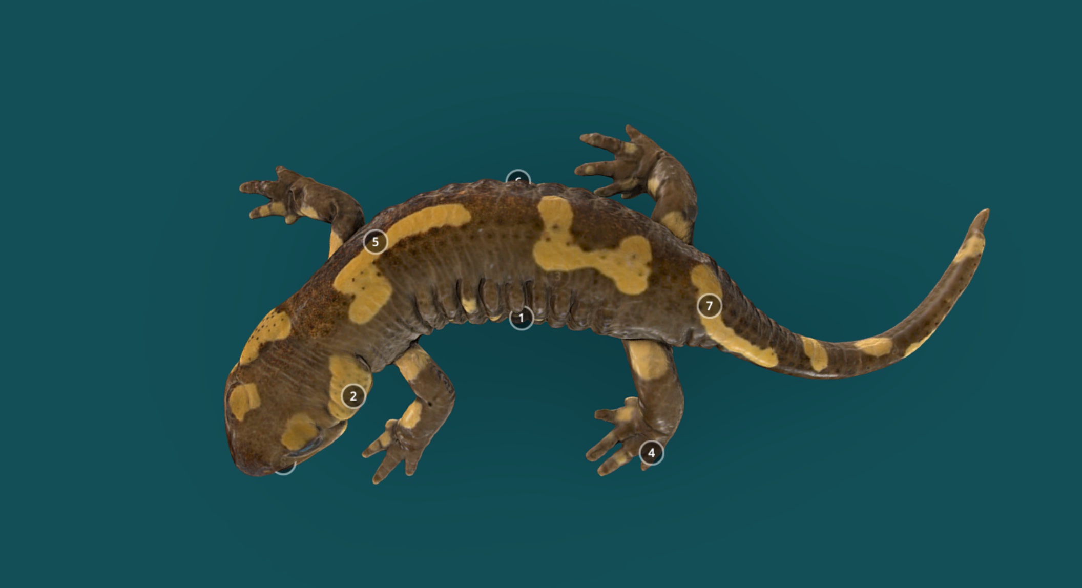
Scanning produces digital information about the geometry and texture of an object that the computer can then render into a 3D, interactive model on your screen. The texture data represent the “look” of the specimen, such as the color or pattern you see on the surface. For example, the texture of a skull scan would include the beige, bone color and any dirt, imperfections, and markings (such as ID number or genus species name) that exist on the surface of the skull. Geometry, on the other hand, includes all shape information about a specimen. This is the actual 3D configuration of the object. A skull’s geometry includes each surface and curve which composes the skull. Structured light scanners capture both types of data with relative speed and accuracy and then affiliated computer software combines them to give you a model that looks impressively like the one sitting in front of you.
Project Goals and Future Aims
My aim with the Oxford Redisplay project was to create accurate, useful, and reusable 3D models for both research and public outreach. As a graduate student with previous digitization and scanning experience in other formats (Micro-CT, primarily), the structured light scanners were new to me, but uniquely fascinating. Even after months of producing scans, I’m impressed each time an object is accurately recreated in three-dimensions on the computer screen. Once created, you can then interact with the model by spinning it around and zooming in on it. I saw a lot of opportunity in this concept, as my experience with morphological research has taught me that virtual specimens are useful for many avenues of academic inquiry. Not only did I expect these scans to have academic utility, I also imagined the role they could play for public outreach and engagement. Virtual models can be easily annotated to include additional descriptive information, which can improve visitor comprehension and interest. I hoped these models could be used for online engagement as well as in the museum, potentially as interactive additions to the new main court displays.

Currently, we are working on the completion of a pilot virtual online tour of the Oxford University Museum of Natural History, which incorporates digital models of new display objects throughout. We are also adding models to websites such as Sketchfab (@morethanadodo) and Morphosource, so that people across the world can view them. I am happy with the progress we have made to-date on this project (somewhere around 200 objects have been scanned and digitized), but there is still plenty of work to complete! Luckily, the public engagement team at OUMNH is seeking funding to continue this project with aims to incorporate 3D models into many future outreach projects.
Conclusion
I think 3D digitization will be important for future biological and museums research alike. Beyond the academic and outreach opportunities it presents, scanning objects allows us to capture a permanent digital record of something. It is like a security blanket, offering a “back-up” copy of objects that are otherwise at risk to the misfortunes of the world (think 2018 National Museum of Brazil fire). Creating and databasing these digital copies can prevent objects from being lost to science. Biologically, extinction rates threaten biodiversity and the organisms we are scanning now, sadly, may not exist in the near future, or have already gone extinct. A digital record will allow future scientists to observe and study extinct specimens.
I think digitization enhances the museum experience and offers many positive outcomes to natural history collections. Importantly, scanning objects at the Oxford University Museum of Natural History improves museum and science accessibility as it allows researchers and visitors alike who otherwise may not be able to afford to travel to the United Kingdom to view OUMNH specimens. Scanning also improves public engagement and outreach, because it creates digital (and ideally, open-access) copies of objects through which people can explore and learn about the world around them.
Taylor West
Taylor West is a PhD candidate in the department of Ecology and Evolution and a member of the Museum Studies Program 2021 cohort at the University of Michigan. She is passionate about digitization within museum collections and spaces, and has recently completed her Museum Studies practicum at the Oxford University Museum of Natural History. When she isn’t scanning specimens, you’ll find Taylor either running or reading (but not simultaneously… yet).
Check out Taylor’s work on the Life as We Know It and The Future in Our Hands exhibits at OUMNH.